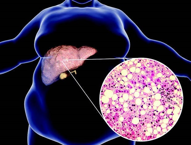
New technology allows for a new way of counting fatty liver cells quickly, accurately and consistently.
Non-alcoholic fatty liver disease (NAFLD) is on the rise in Singapore, not unlike in the West where diet and lifestyle play important roles. NAFLD, associated with diabetes and obesity, can lead to liver cirrhosis or scarring and hardening, and liver cancer.
When signs of the disease appear, a biopsy is sometimes necessary to gauge the accumulation of fatty cells, inflammation damage and fibrous scar tissue in the liver. But the conventional method of quantifying fatty cells sometimes may not be able to determine the stage of the disease.
The current technique where the pathologist peers through a microscope to assess liver fat in a tissue sample can only work out broad categories — 0-5 per cent (no liver disease), 5-33 per cent (mild), 33-66 per cent (moderate) or more than 66 per cent (severe). Although most biopsies can be clearly graded, it is less obvious in some cases. If the amount of fatty cells look like it might be around 65-66 per cent, is it in the moderate or severe stage? And how should the patient be treated?
Following advances in technology, a team of Singapore General Hospital (SGH) clinicians has developed an automated test that in a study was found to be highly accurate and consistent in its analysis. The test involves an optical microscopy technique known as secondary harmonic generation (SHG) microscopy to quickly produce a digital image of the tissue. A computerised algorithm or formula developed by the team is then used to detect and count the fat cells.
“This study is important as it shows for the first time that SHG microscopy can be used to assess liver fat. It can thus be an additional tool to help standardise the quantitative analysis of liver steatosis,” said Associate Professor Jason Chang, Senior Consultant and Head, Department of Gastroenterology and Hepatology, SGH.
The current gold standard is subjective as it relies on pathologists’ experience and knowledge, whereas the new method standardises and gives its readings in precise percentages. This is important especially in trials where accurate quantification is crucial, Prof Chang said.
Putting into practice
In the study, the team studied liver biopsies from 86 patients diagnosed with NAFLD and/or chronic hepatitis B at SGH between 2006 and 2016. They compared liver fat assessment using the new SHG tool and by three expert pathologists. No significant differences were found between the two, demonstrating the accuracy of the automated SHG method.
As the study was relatively small with sample tissues from just one centre, future studies are being planned to involve larger cohorts of participants from multiple centres, said Dr George Goh, Senior Consultant, Department of Gastroenterology and Hepatology, SGH, and the study’s first author.
At the same time, the team is working with DxD Hub, a unit of A*Star, to implement the algorithm into clinical practice at SGH, as well as international researchers to use SHG to measure other features of fatty liver disease.
Although “this assistive tool helps the pathologist quantify the liver fat in a more precise manner, removing inter- and intra-observer variations”, it won’t replace the pathologist just yet, said Dr Leow Wei Qiang, Consultant, Department of Anatomical Pathology, SGH. The algorithm may have been designed to pick out features specific to fatty liver, but it cannot tell if a biopsy is of a fatty liver tissue or something else in the first place.

The Quantification of hepatic steatosis in chronic liver disease using novel automated method of second harmonic generation and two-photon excited fluorescence study was published in Scientific Reports, a leading peer-reviewed science journal, on 27 February 2019. Other team members are Professor Tan Chee Kiat of the Department of Gastroenterology and Hepatology, SGH; Associate Professor Tony Lim and Dr Wan Wei Keat of the Department of Anatomical Pathology, SGH; and Dr Shen Liang of the Biostatistics Unit, Yong Loo Lin School of Medicine, NUS.
The team’s breakthrough in measuring liver fat arose from an earlier collaboration with medical technology firms HistoIndex Pte Ltd and DxD Hub using SHG to stage liver scarring (fibrosis) in patients with NAFLD.


 Get it on Google Play