


reduces the risk of development and progression of diabetic retinopathy in both type 1 and type 2 diabetes.
Diabetic Retinopathy: Updates in Detection and Treatment
By: Dr Gavin Tan, Consultant, Surgical Retina Department, Retina Centre, Singapore National Eye Centre
Diabetes affects approximately 1 in 12 Singaporeans aged 18 to 69 years, and in absolute terms there are more than 300,000 persons with this chronic debilitating disease. This figure is likely to increase further because of Singapore’s ageing population and increasing prevalence of diabetes risk factors, such as obesity and sedentary practices.
Diabetic Retinopathy (DR) is the most common microvascular complication of diabetes and the leading cause of vision loss in working-aged adults worldwide. Diabetic retinopathy is characterised by changes in the retina including haemorrhages, microaneurysms, arteriolar and venular dilatation. These have a progressive nature eventually leading to areas of retinal non-perfusion, increased vasopermeability with retinal oedema and exudates, and pathologic proliferative of intraocular blood vessels resulting in haemorrhage, tractional retinal detachment or neovascular glaucoma all of which contribute to visual impairment and blindness.
EPIDEMIOLOGY OF DIABETIC RETINOPATHY
The Singapore Epidemiology of Eye Diseases (SEED) study group consisted of three population-based eye studies that examined the epidemiology of eye diseases in the three major ethnic groups, the Malays (SiMES), the Indians (SINDI) and the Chinese (SCES). The overall age-standardised prevalence figures of any DR, and other more severe stages of DR such as diabetic macular oedema (DME), and vision-threatening retinopathy (VTDR), were found to be 28.2%, 7.6%, and 7.7% respectively, among persons with diabetes.
Comparing the prevalence of DR between ethnic groups, Indians with diabetes were found to have a high prevalence of DR and DME (Table 1). Even after adjusting for known risk factors for DR and socioeconomic status, Indians had a 50% higher risk of diabetic retinopathy (p=0.002) compared to Chinese with diabetes.

Vision loss from DR is largely related to two late-stage conditions: diabetic macular oedema (DME) and proliferative diabetic retinopathy (PDR). However, the number of people with PDR has markedly reduced over the last three decades due to improved systemic management of diabetes, early detection of retinopathy through screening programs, and timely laser treatment.
Thus, DME has now overtaken PDR as the most frequent cause of visual impairment among persons with diabetes in developed countries. In our own study population, 7.6% of diabetics had DME compared with 3.8% for PDR.
SCREENING FOR DIABETIC RETINOPATHY
Regular retinal examination is the cornerstone of effective diabetes management aiming to detect DR before it causes visual loss so that effective treatment can be given.
Studies in Sweden, Iceland and the United Kingdom have found as much as a two-thirds reduction of diabetes-related blindness after implementation of a national diabetic retinopathy screening program. The Singapore Ministry of Health (MOH) guidelines recommend that people with diabetes be screened annually for diabetic eye disease, or referred for tertiary care for those with evidence of the disease.
Retinal photography is the main screening tool used for diabetic retinopathy, since it would not be feasible to have all 300,000 diabetics visit an ophthalmologist annually. The diabetic retinal photography program in Singapore was implemented in the polyclinics in 1990. These early cameras provided a single-field printed photograph of posterior pole of the retina, which was read by family physicians.
The Singapore Integrated Diabetic Retinopathy Programme (SiDRP) was implemented as an innovative telemedicine model to optimise screening for DR at the primary care level. The SiDRP uses digital cameras to capture two-field retinal photographs for each eye which are transmitted electronically to a reading centre.
The images are then graded by a centralised team of trained and accredited technicians, and reports are generated within an hour and sent to the patients’ clinician who will subsequently make appropriate referrals to tertiary eye care based on the findings and recommendations (Figure 1).

The SiDRP program started out initially as a pilot project in Outram and Bukit Merah Polyclinic in 2010 with one reading centre at Singapore National Eye Centre (SNEC) / Singapore Eye Research Institute (SERI). It has subsequently been expanded to all 18 clinics nationwide with two reading centres, one at SNEC and the other at the National Healthcare Group (NHG) Eye Institute in 2016.
In addition, today, we provide telemedicine diabetic retinopathy grading service for other primary healthcare facilities including the six community health centres, family medicine centres, Endocrinology clinics at restructured hospitals [Changi General Hospital (CGH), KK Women’s and Children’s Hospital (KKH), National University Hospital (NUH) and Singapore General Hospital (SGH)] and other organisations such as the Diabetic Society of Singapore and Singapore Anti-Tuberculosis Association (SATA), which provide access to patients who receive their diabetes care outside of the polyclinic network.
CHANGES IN THE TREATMENT PARADIGM
Systemic Treatment
The most effective treatment for diabetic retinopathy is prevention and this involves the optimisation of systemic risk factors. Hyperglycaemia is the key initiator in the pathogenesis and development of diabetic retinopathy.
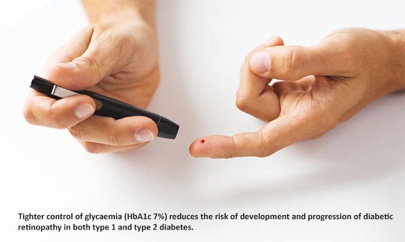
The Diabetes Control and Complications Trial (DCCT) and the United Kingdom Prospective Diabetes Study (UKPDS) are the key studies providing definitive evidence that tighter control of glycaemia (HbA1c 7%) reduces the risk of development and progression of diabetic retinopathy in both type 1 and type 2 diabetes, with each percent reduction in HbA1c (e.g. from 9% to 8%) lowering the risk of retinopathy by 30% to 40%.
Recent analysis from the UKPDS and DCCT have suggested that the protective effect of intensified blood glucose control, especially early in disease, has a sustained effect over time. This ’metabolic memory‘ effect persists even if glycaemic control is less intensive later in the course of disease.
In the UKPDS, at 10 years after the end of the trial, although the between group differences in glycated haemoglobin levels were lost after the first years, in the intensively treated group, the relative risk reduction persisted at 24% for microvascular disease compared with the less intensive group.
Blood pressure control remains the second key pillar of DR prevention with each 10 mmHg increase in systolic blood pressure associated with an approximately 10% excess risk of early diabetic retinopathy and a 15% excess risk of proliferative retinopathy, and in the UKPDS, tighter blood pressure control reduced the risks of retinopathy progression by about one-third, visual loss by half, and need for laser treatment by one-third in people with type 2 diabetes.
Clinical trials have shown that renin-angiotensin system inhibitors may reduce the incidence and progression of diabetic retinopathy beyond their blood pressure-lowering effects compared with other anti-hypertensive drugs.
Observational studies support a role for dyslipidaemia in the pathogenesis of diabetic retinopathy. The Fenofibrate Intervention and Event Lowering in Diabetes (FIELD) trial showed that fenofibrate, a lipid-modifying agent, reduced the need for laser treatment of vision-threatening diabetic retinopathy by 31% in patients with type 2 diabetes over five years.
The Action to Control Cardiovascular Risk in Diabetes (ACCORD) study reported a 40% reduction in the odds of having progression of retinopathy over four years afforded by fenofibrate combined with simvastatin, compared with simvastatin alone, further supporting the efficacy of fenofibrate in reducing the progression of diabetic retinopathy. Importantly, these effects were independent of the changes to the lipid profile.
In Singapore, the MOH diabetes clinical guidelines recommend consideration be given to the use of fenofibrate to retard diabetic retinopathy (level B evidence). The Health Sciences Authority also recently approved micronised fenofibrate (Lipanthyl, Abbott) for the indication of reduction in the progression of diabetic retinopathy in patients with type 2 diabetes and existing diabetic retinopathy.
Management of DME and DR
DME is characterised by exudative fluid accumulation at the macula, the area of the retina responsible for sharp central vision. DME can occur at any stage of DR, although the risk of DME increases with increasing DR severity. The clinical signs of DME include hard exudates with microaneurysms and blot haemorrhages within one disc diameter of the centre of the macula (Figure 2).

Optical Coherence Tomography (OCT) is a new imaging modality using low coherence interferometry to provide a noncontact non-invasive optical ’biopsy‘ of the retina, allowing objective qualitative and quantitative assessment of DME.
OCT presents data on retinal thickness (Figure 2), and can also be used to qualitatively identify DME based on morphological features, such as intraretinal fluid, intraretinal cysts, and subretinal fluid. OCT is better than clinical slit lamp biomicroscopy and fundus photography for the assessment of mild DME and has become the standard for diagnosis and monitoring of DME.
Since the ETDRS study was published in the 1990s, macular focal/grid laser therapy has been the gold-standard treatment for DME. In the past decade, Vascular endothelial growth factor (VEGF) has been found to be a key mediator of DR and DME. With the emergence of anti-VEGF drugs, there has been a major paradigm shift in the ocular management of DME.
Monthly intraocular injections of drugs targeting VEGF have been shown in a number of randomised controlled trials to conclusively result in better visual outcomes than that of traditional laser therapy for centre involving DME.
There are currently three commonly used anti-VEGF drugs with efficacy proven in clinical trials for treating DME, ranibizumab, aflibercept and bevacizumab, which are injected trans-sclerally (trans pars plana) into the vitreous cavity with a small gauge needle. The former two drugs were specifically designed and approved for ocular use, while the latter is a cancer drug used on an off-label basis for the treatment of retinal vascular diseases.
Although, the use of bevacizumab is off-label, it is one of the most widely-used anti-VEGF drugs globally for ocular diseases, because it has proven efficacy for DME and age-related macular degeneration in randomised control trials and it is far cheaper than the other alternatives.
Multiple monthly injections are usually required to treat the oedema, and laser can be used as an adjuvant treatment (Figure 3). A recent randomised control trial compared the efficacy of the three commonly used anti-VEGF agents for DME and found that at one year and two years, all three drugs were effective with improvement of visual acuity from baseline and a decreased number of injections in year 2. Among eyes with worse vision at the start of the study, aflibercept had superior two-year visual outcomes compared with bevacizumab.
Anti-VEGF are an effective treatment for DME – however, the cost of treatment, which is up to thousands of dollars per injection, and the burden of monthly intravitreal injections remain significant. Laser will still have a role in the treatment of non-centre involving DME and as an adjuvant to intravitreal anti-VEGF injections.
There is recent evidence that anti-VEGF injections can reduce the progression of DR and even lead to regression of DR when used in the treatment of DME. A randomised clinical trial comparing pan retinal photocoagulation (PRP) and intravitreal ranibizumab found that among eyes with proliferative diabetic retinopathy, treatment with ranibizumab resulted in visual acuity that was non-inferior to (not worse than) PRP treatment at two years.
However, the ranibizumab group required a median of 10 injections over two years compared with one to three sessions of laser in the PRP group, and there is no evidence to when anti-VEGF injection can be stopped, and what will happen to the DR after stopping injections. The significant cost and burden of such intravitreal injections make it currently unlikely that it will replace PRP as the standard of care.

CONCLUSION
Diabetic retinopathy is an important cause of visual loss in Singapore and affects 30% of those with diabetes. Prevention of DR with systemic control remains key and the addition of fenofibrate may play a role.
Screening for DR in diabetics is essential to prevent visual loss. The SiDRP, a new telemedicine DR screening program should improve access to DR screening across Singapore.
Intraocular injections of anti-VEGF drugs have emerged as the gold standard for the treatment of centre involving DME, however, the cost and burden of monthly treatment is a significant public health issue and a barrier to optimal care.
GPs can call for appointments through the GP Appointment Hotline at 6322 9399.
Dr Gavin Tan is a Consultant in the Surgical Retina Department and Retina Centre of the Singapore National Eye Centre (SNEC). He completed multiple vitreo-retinal fellowships at SNEC, the Byers Eye Institute at Stanford University and Jules Stein Eye Institute at the University of California Los Angeles (UCLA). He is actively involved in academic research and leads multiple clinical trials at the Singapore Eye Research Institute (SERI). His major clinical and research interest is in the field of diabetic retinopathy.
Stay Healthy the Easy Way
Get trusted health advice, offers and more.

Get the Health Buddy App
 Get it on Google Play