


One of the very common conditions encountered by clinicians in their outpatient setting is trigger finger. As prevalent as 2% in the general population, the prevalence approaches 20% in diabetic patients.1
Recognised as a pathological condition of the flexor tendons, it was first described by the French physician Notta in 1850.2 Saldana later refined this in 2001 as a pathologic disproportion between the volume of the flexor tendon sheath and its contents.3
Pathophysiology, Symptoms and Signs
Trigger finger results from a narrow tunnel created from an inflamed and thickened pulley. This leads to the obstruction of the free and smooth gliding of the tendons resulting in locking, clicking, reduced excursion or no excursion at all.4
Eventually, patients present with trigger symptoms which may vary from the initial stages of pain over volar metacarpophalangeal joint with or without evidence of locking or clicking, progressing to reduced movement of the finger joints and finally to a fixed locked finger in flexion.
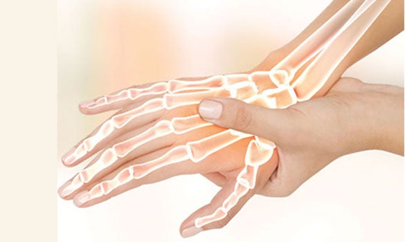
The first annular pulley (A1) is usually affected in most cases of trigger fingers (Figure 1), though cases of triggering have been reported at the second (A2) and third (A3) annular pulleys, as well as the palmar aponeurosis.

Microscopic examination of normal A1 pulleys demonstrates an amorphous extracellular matrix, including chondrocytes, coating the pulley’s entire innermost layer. Pathological changes involving A1 pulleys in trigger fingers show areas of extracellular matrix loss along with chondrocyte proliferation and type III collagen production. This fibrocartilagenous metaplasia is thought to result from the repeated friction and compression between the flexor tendon and the corresponding inner layer of the A1 pulley.6
Based on the symptoms, trigger finger can be divided into 4 grades in accordance to Green’s classification7:
Table 1
Grades of trigger and associated signs and symptoms
| Grade | Symptoms and Signs |
|---|---|
| Grade 1 (pre-triggering) | Pain; history of catching, but not demonstrable on physical examination; tenderness over the A1 pulley |
| Grade 2 (active) | Demonstrable catching, but the patient can actively extend the digit |
| Grade 3 (passive) | Demonstrable catching requiring passive extension (grade 3A) or inability to actively flex (grade 3B) |
| Grade 4 (contracture) | Demonstrable catching with a fixed flexion contracture of the PIP joint |
A study conducted at Singapore General Hospital by Choudhury and Tay8 of patients with trigger finger showed a female preponderance with a female to male ratio of 2:1. The majority of cases were patients in the 6th decade. The middle finger was most commonly involved, followed by the ring finger. The least involved digit was the little finger. The most common grade at presentation was grade 2 (41%) and the least common grade was grade 4 (2%).
What Can We Do for Our Patients?
Currently, the different treatment modalities include a combination of topical non-steroidal anti-inflammatory drugs (NSAIDs), occupational therapy, splinting, corticosteroid injections and trigger finger release.
When to Refer to a Hand Surgeon?
Some patients are reluctant to endure an invasive and painful procedure like an injection. These patients can be referred to hand surgeons at a tertiary level hospital for combination therapy. Patients having recurrence after the injection or those who have already had two injections without resolution of their symptoms are more likely to benefit from surgery and would be the ideal candidates for a referral to a hand surgeon.
Corticosteroid Injections (H & L)
Multiple studies have been done on the efficacy of corticosteroid injection. In general, long-term relief can be achieved in 60 to 92% of affected digits. The efficacy of the injection drops in patients with longstanding disease (more than 6 months duration), diabetes mellitus and multiple digit involvement as it is unable to reverse the changes of fibrocartilagenous metaplasia in the A1 pulley. 60% of trigger digits receiving corticosteroid injection eventually require surgery.6
Nevertheless, H & L injections remain an extremely viable option for patients in the primary care setting as it can be administered immediately with relief of symptoms within a week, curtailing the need of long periods of splinting and therapy sessions and waiting for an operation date.
Traditionally, corticosteroid injection is injected into the flexor tendon sheath just over the metacarpal head. A 1 ml mixture of equal proportions of triamcinolone and 1% Lignocaine or 0.5% Marcaine is injected under aseptic conditions. The mixture is drawn into a 3 ml or 5 ml syringe and then administered via a 23G needle. The syringe is held like a pen and the needle is introduced through the skin at an angle of 45° until it pierces the flexor tendons. The needle is then gradually withdrawn whilst pressure is applied over the plunger (Figure 2).

Once a ‘give’ is felt, 1 ml of the mixture is injected into the flexor sheath which sometimes can be a felt as a fluid thrill along the volar aspect of the finger. If symptoms do not resolve after the first injection, or recur afterwards, a second injection is typically half as likely to succeed as the initial treatment.
Some authors have found that the intrasynovial injection to be painful9 and at times associated with injury to the tendon. 10 The question then arises that is it always necessary to inject into the sheath? It has been shown that the injectable load delivered just in the subcutaneous layer above the pulley appears to be as efficacious as a cumbersome intrasynovial injection.11
Where complications are concerned, tendon rupture is fairly rare. It has been associated with multiple corticosteroid injections, leading us to the rule-of-thumb of administering a maximum of two injections per finger. Other complications include dermal atrophy, fat necrosis, skin hypopigmentation, transient elevation of serum glucose in diabetic patients and infection. None of the cases in the local study had any injection-related complications.8
Combination Therapy
From the local study8, the most common initial treatment administered was combination therapy (Figure 3) consisting of trigger, splint occupational therapy sessions and topical NSAIDs (Figure 4). This was given mainly for grade 1 and 2 triggers.
Occupational therapy sessions involved laser treatment, fluidotherapy and tendon gliding exercises requiring an average of 5 occupational therapy sessions over a mean period 2 months.
The goal of splinting was to prevent the friction caused by flexor tendon movement through the affected A1 pulley until the inflammation there resolved. The combination of laser therapy and NSAIDs enhances the effect of the splint, thus making it a desirable option for patients with less severe trigger symptoms who are more adverse to the needle and scalpel.


Open Trigger Finger Release
The surgical release of trigger finger is a very common procedure bearing excellent results12 with 97% complete resolution of triggering. Release is done by dividing the A1 pulley which is the usual site of the stenosis.
In the local study8, all the patients who underwent surgery had successful resolution of their symptoms. None of these patients required further operations. One patient, however, had a wound breakdown which healed eventually with dressings. There were no other complications in patients who underwent surgery.
Conclusion
Trigger fingers are a fairly common disorder of the digits. There is an increased prevalence amongst diabetic patients. Triggering represents a painful nuisance hampering daily activities, dampening efficiency at the workplace and at times translates to prolonged periods of absence from work.
With the availability of a simple, effective and safe procedure like the corticosteroid injection, family physicians serving the community can add to their armamentarium an early solution to the patient’s painful digit.
GPs can call for appointments through the GP Appointment Hotline at 6321 4402 for more information.
By: Dr Darryl Chew, Consultant, Department of Hand Surgery, Singapore General Hospital and Dr Muntasir Mannan Choudhury, Registrar, Department of Hand Surgery, Singapore General Hospital
Dr Darryl Chew is a Consultant with the Department of Hand Surgery at the Singapore General Hospital. Hand trauma forms the bulk of his caseload at SGH. His sub-specialty interest is in the paediatric hand and sees children presenting with hand problems at KK Women’s and Children’s Hospital.
Dr Muntasir Mannan Choudhury is a Registrar with the Department of Hand Surgery at the Singapore General Hospital. His areas of interest include nerve, microsurgery and reconstruction of the hand.
References
1. Fitzgibbons PG, Weiss AP. Hand manifestations of diabetes mellitus. J Hand Surg Am. 2008 May- Jun;33(5): 771-5.
2. A. Notta. Research about a particular affection of the tendons of the hand, characterized by the development of a nodosity on the trajectory of the flexor tendons of the digits that impedes their movements. Archives generales de medecine, 4 (1850), pp. 142–161.
3. Saldana MJ. Trigger digits: diagnosis and treatment. J Am Acad Orthop Surg. 2001, 9(4): 246-252.
4. Ryzewicz M, Wolf JM. Trigger digits: principles, management, and complications. J Hand Surg Am.2006 Jan; 31(1): 135-146.
5. Andrew Chin, Teoh Lam Chuan. Painful Conditions of the Hand. Trigger digits. P 39, Chap 5.
6. Sbernardori MC, Mazzarello V, Tranquilli-Leali P. Scanning electron microscopic findings of the gliding surface of the A1 pulley in trigger fingers and thumbs. J Hand Surg [Br] 2007;32:384.
7. Wolfe SW, Hotchkiss RN, Pederson WC, Kozin SH, Tendinopathy, in: Green’s Operative Hand Surgery, 6th edition, Churchill Livingstone, Chap. 62, p. 5, 2011.
8. Choudhury MM, Tay SC. Prospective Study on the Management of Trigger Finger. Hand Surgery, Vol. 19, No. 3 (2014) 393–397.
9. K. Kazuki et al. Clinical outcome of extrasynovial steroid injection for trigger finger; Hand Surgery, Vol. 11, Nos. 1 & 2 (2006) 1–4.
10. S. Jianmongkol et al. Intra-tendon sheath injection for trigger finger: the randomized controlled trial. Hand Surgery, Vol. 12, No. 2 (2007) 79–82.
11. Taras et al. Corticosteroid injections for trigger digits: Is intrasheath injection necessary? J Hand Surgery 1998; 23A: 717-722.
12. Turowski GA, Zdankiewicz PD, Thomson JG. The results of surgical treatment of trigger finger. J Hand Surg 1997;22A:145–149.

Stay Healthy the Easy Way
Get trusted health advice, offers and more.

Get the Health Buddy App
 Get it on Google Play