


3D mammograms and contrasted mammograms: what are they, and why do general practitioners need to know about them? Although you may not encounter these imaging techniques often in your daily practice, you may occasionally have patients asking about them, particularly given our increasingly health-conscious population.
Patients participating in the national breast cancer screening programme who have abnormal findings on initial screening mammograms will often be recalled for further assessment, which entails usage of newer techniques such as 3D mammograms and contrasted mammograms.
This article provides an overview of their indications, utility and limitations.
1. DIGITAL BREAST TOMOSYNTHESIS
WHAT IS DIGITAL BREAST TOMOSYNTHESIS?
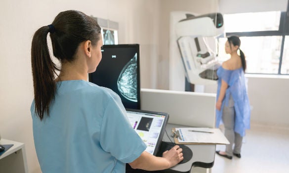
Digital breast tomosynthesis (DBT), also known as 3D mammogram, is a type of mammography that uses X-rays to produce detailed three-dimensional images of the breast tissue.
How does it work?
It involves multiple projections acquired across an arc that are reconstructed into a series of stacked images. The tomosynthesis data set can then be used to recreate two-dimensional images using synthetic mammography (SM), producing images almost equivalent to those of the traditional full-field digital mammography (FFDM).1
Hence, in a DBT study, you will get a series of stacked images of each breast in two views and a set of synthesised mammogram which is almost of traditional mammogram equivalence.
WHEN AND WHY DO WE USE DBT?
The stacked images from DBT facilitate visualisation, localisation and characterisation of a lesion.
Further assessment of asymmetric density on screening mammogram
Some patients may be recalled for assessment of asymmetric density present on the screening mammogram. The multiple sections and additional angles available with DBT imaging often allow the resolution of areas of normal superimposed tissue that may present as summations or higher density areas in FFDM.
If the supplementary ultrasound also did not reveal any significant abnormalities, patients can usually be discharged back to routine rescreening without further investigation or biopsy.
Detecting mammographic architectural distortion
The beauty of DBT lies in its ability to comprehensively assess mammographic architectural distortion, the most common missed abnormality in interval cancers. It has also been shown to improve the detection of such distortion.
Figure 1A shows an example of a patient with extremely dense breasts, who was recalled for assessment of possible architectural distortion in the right breast. The area of distortion was incredibly subtle on the screening mammogram. In fact, it was missed by one reader, but was depicted clearly on DBT.
Supplementary ultrasound examination showed a corresponding irregular hypoechoic lesion which was suspicious in nature, and was biopsied under ultrasound guidance (Figure 1B). Histology yielded invasive ductal carcinoma with lobular differentiation.

Lower recall rates and increased cancer detection rates
Some countries with historically high breast cancer screening recall rates have managed to significantly lower their recall rates and improve specificity after replacing FFDM with DBT imaging as a first-line screening tool (FFDM remains as the first-line screening tool in Singapore).
Prospective and retrospective observational studies have reported a 15% to 63% decrease in recall rate.1-5
Other studies have demonstrated an increase in cancer detection rate (CDR) after employing DBT as a screening tool. In particular, there was a higher pick-up rate of both smaller as well as lower-grade malignancies.1-3
That said, its long-term impact on patient survival remains uncertain as breast cancer survival benefits are difficult to prove without extended follow-up and/ or randomised controlled trials.
DBT use in Singapore
DBT is not currently used as a screening tool in Singapore. It is associated with longer acquisition and interpretation times owing to the larger image set.
There is also a slight increase in radiation dose with DBT relative to FFDM.
Currently, it serves as an adjunct problem-solving imaging tool in the hospital setting to assess abnormal breast findings and assist in lesion-localisation for biopsy.
Ongoing research to reduce DBT reading time and improve its imaging performance may change the status quo.1
COMMON QUESTIONS THAT PATIENTS MAY ASK |
|---|
HOW MUCH MORE RADIATION DOES DBT HAVE COMPARED TO A NORMAL MAMMOGRAM? In a review by Svahn et al., the authors concluded that while using DBT plus FFDM increases radiation dose by a factor of up to 2.25 compared with FFDM alone, using SM to replace the FFDM portion can bring the radiation dose to a level comparable to that of FFDM alone.4 According to Skaane et al., the dose with DBT plus SM is reported to be just 19% higher than that with FFDM alone. The use of SM in place of FFDM with DBT pro-vides similar outcomes while reducing radiation dose.5 In summary, modern-day DBT imaging has a similar or just slightly higher radiation dose as compared to a normal mammogram. I HAVE HEARD ABOUT THIS ‘SPECIAL MAMMOGRAM’. AM I ABLE TO GO FOR IT? DBT is only indicated in certain specific cases, which first requires an initial evaluation by a breast surgeon or radiologist. This facility is only available in hospitals and not in government polyclinics. |
2. CONTRAST-ENHANCED SPECTRAL MAMMOGRAM
WHAT IS A CONTRAST-ENHANCED SPECTRAL MAMMOGRAM?
Contrast-enhanced spectral mammogram (CESM) provides additional information beyond a conventional mammogram and may aid earlier detection of some cancers. It is a recent development in digital mammography that utilises dual-energy exposure following the injection of an iodinated contrast agent.5-6
How does it work?
Typically, an iodinated contrast agent (1.5 ml/kg of body weight) will be administered to the patient via intravenous injection. Two minutes later, standard mediolateral oblique (MLO) and craniocaudal (CC) projections will be undertaken on each breast.
Each projection will receive two exposures, one with a low energy (around 30 kVp) and the other with a high energy (around 45 kVp). A specific dual-energy image recombination algorithm is used to subtract the low- and high-energy images, resulting in an ‘iodine uptake map’.
Hence, there will be two sets of images in a CESM study:
A set of low-energy images, which looks like a conventional mammogram
A set of recombined images, which displays contrast medium uptake
The entire process usually takes about 30 minutes.6-7
CESM, likened to MRI breasts, leverages on tumoural neoangiogenesis.8,9 It provides both morphological information, similar to a routine mammogram, and functional information about a lesion’s enhancement pattern. Several studies have demonstrated the superior sensitivity and low false positive rates of CESM, with studies exploring its potential as an alternative to MRI breasts in breast cancer staging and problem solving.6-8
CESM use in Singapore
This imaging technique has been available in Singapore for the last five to eight years. We have found it relatively easy to adopt in a hospital setting, and it has been helpful in our clinical practice.
WHEN AND WHY DO WE USE CESM?
Indications
CESM is indicated:
- For the evaluation of an ultrasound-occult asymmetric density
- For delineation of tumour extent
- As a problem-solving tool in complicated cases
Contraindications
Contraindications for CESM are identical to contrast-enhanced CT scans, which include:
- Renal impairment
- Contrast allergy
- Asthma
- Pregnancy (due to radiation) – the radiation dose of CESM relative to mammogram reportedly ranges from 1.06 to 1.437
Improving diagnostic accuracy and care
The addition of CESM to our workflow has helped improve diagnostic accuracy and patient care. As an example, the initial assessment of a patient with abnormal findings in the left breast showed a suspicious mass and an indeterminate left axillary lymph node. They were biopsied under ultrasound guidance and proven to be invasive breast cancer and metastatic left axillary adenopathy respectively (Figure 2A).
The addition of CESM into the workflow enabled the detection of an additional enhancing lesion in the right breast (Figure 2B) which was not visible on routine mammogram and ultrasound. This enhancing lesion in the right breast, detected only on CESM, was also subsequently proven to be invasive breast cancer.
According to reports, the false positive rate of CESM is
comparable to or even lower than that of MRI breasts.9 There is ongoing research into CESM screening and
CESM-guided biopsy, with this imaging technique
having generated a fair bit of excitement among the
breast imaging community.
COMMON QUESTIONS THAT PATIENTS MAY ASK |
|---|
WILL THERE BE AN ALLERGIC REACTION RELATED TO CONTRAST ADMINISTRATION IN CESM? HOW IS IT MANAGED?
|
CONCLUSION
DBT and CESM are relatively new breast imaging techniques that have recently cemented their places in clinical practice. They are practical tools that we use in the hospital setting, and a brief understanding of them may help in communication with your patients and for continuity of care.
REFERENCES
Chong A, Weinstein SP, McDonald ES, Conant EF. Digital Breast Tomosynthesis: Concepts and Clinical Practice. Radiology. 2019;292(1):1-14.doi:10.1148/radiol.2019180760
Conant EF, Beaber EF, Sprague BL, et al. Breast cancer screening using tomosynthesis in combination with digital mammography compared to digital mammography alone: a cohort study within the PROSPR consortium. Breast Cancer Res Treat. 2016;156(1):109-116. doi:10.1007/s10549-016-3695-1
Sharpe RE, Venkataraman S, Phillips J, et al. Increased Cancer Detection Rate and Variations in the Recall Rate Resulting from Implementation of 3D Digital Breast Tomosynthesis into aPopulation-based Screening Program. Radiology. 2016;278(3):698-706. doi:10.1148/radiol.2015142036
Svahn TM, Houssami N, Sechopoulos I, Mattsson S. Review of radiation dose estimates in digital breast tomosynthesis relative to those in two-view full-field digital mammography. The Breast. 2015;24(2):93-99. doi:10.1016/j.breast.2014.12.002
Skaane P, Bandos AI, Gullien R, et al. Prospective trial comparing full-field digital mammography (FFDM) versus combined FFDM and tomosynthesis in a population-based screening programme using independent double reading with arbitration. Eur Radiol. 2013;23(8):2061-2071. doi:10.1007/s00330-013-2820-3
James JJ, Tennant SL. Contrast-enhanced spectral mammography (CESM). Clinical Radiology. 2018;73(8):715-723. doi:10.1016/j.crad.2018.05.005
Dromain C, Vietti-Violi N, Meuwly JY. Angiomammography: A review of current evidences. Diagnostic and Interventional Imaging. 2019;100(10):593-605. doi:https://doi.org/10.1016/j.diii.2019.01.011
Tagliafico AS, Bignotti B, Rossi F, et al. Diagnostic performance of contrast-enhanced spectral mammography: Systematic review and meta-analysis. The Breast. 2016;28:13–19
Elżbieta Łuczyńska, Sylwia Heinze-Paluchowska, Edward Hendrick, et al. Comparison between Breast MRI and Contrast-Enhanced Spectral Mammography. Medical Science Monitor. 2015;21:1358-1367. doi:10.12659/MSM.893018
Houben IPL, Van de Voorde P, Jeukens CRLPN, et al. Contrast-enhanced spectral mammography as work-up tool in patients recalled from breast cancer screening has low risks and might hold clinical benefits. European Journal of Radiology. 2017;94:31-37. doi:10.1016/j.ejrad.2017.07.004
Modi K, Padala SA, Gupta M. Contrast-Induced Nephropathy. In: StatPearls. StatPearls Publishing; 2022. Accessed February 1, 2023. http://www.ncbi.nlm.nih.gov/books/NBK448066/
Hossain MA, Costanzo E, Cosentino J, et al. Contrast-induced nephropathy: Pathophysiology, risk factors, and prevention. Saudi Journal of Kidney Diseases and Transplantation. 2018;29(1):1. doi:10.4103/1319-2442.225199
Cope LH, Drinkwater KJ, Howlett DC. RCR audit of compliance with UK guidelines for the prevention and detection of acute kidney injury in adult patients undergoing iodinated contrast media injections for CT. Clinical Radiology. 2017;72(12):1047-1052. doi:10.1016/j.crad.2017.07.002
Dr Amanda Liew graduated from the University of Edinburgh with honours in 2014 and became a Fellow of the Royal College of Radiologists in 2018. She attained her specialist accreditation in diagnostic radiology in 2021. She is currently an Associate Consultant at the National Cancer Centre Singapore, and her special interests are in breast imaging, breast cancer research and education.
GPs can call the SingHealth Duke-NUS Breast Centre for appointments at the following hotlines or click here to visit the website:
Singapore General Hospital: 6326 6060
Changi General Hospital: 6788 3003
Sengkang General Hospital: 6930 6000
KK Women’s and Children’s Hospital: 6692 2984
National Cancer Centre Singapore: 6436 8288
Stay Healthy the Easy Way
Get trusted health advice, offers and more.

Get the Health Buddy App


Follow HealthXchange

