


More than 110 years ago, German physics professor Wilhelm Conrad Rontgen took the world’s first photograph of a human body part using X-rays – that of his wife’s hand. The professor referred to the form of radiation as X, meaning it was an unknown type of radiation, and went on to receive the Nobel Prize in Physics for his discovery.
Now X-ray machines are ubiquitous in hospitals and clinics, as are other medical diagnostic tools such as ultrasound and CT (computed tomography) scanners. With technological advances over the years, the devices have become increasingly sophisticated, leading to early detection of illnesses and speedier diagnosis with greater accuracy.
In obstetrics, the ultrasound is the method of choice to examine the foetus as it is safe for the baby. Ultrasound uses sound waves to produce an image.
Dr Terence Kee, a proud father-to-be, speaks with wonderment about the ultrasound scans that his wife has had of their baby. “It gave me a sense of close bonding with the baby and made pregnancy more real. The ultrasound also provided a sense of comfort that the pregnancy was OK,” he said.
“Now the scanners are so marvellous, the images are very clear. I can see the double eyelids on my baby,” he enthused. “When I was a medical student in the 1990s, I couldn’t see such details.” Dr Kee, who works in one of the local hospitals, added.
Cardiologist Dr Chandana Samaranayake notes that technical advances enables the production of three-dimensional (3D) ultrasound images of the foetus. In the past, only two-dimensional pictures were possible.
Dr Chandana has been working at Philips Medical Systems at Philips Electronics Singapore for the past six years. Philips is among the major suppliers of diagnostic imaging and patient monitoring devices.
“With 3D ultrasound, doctors can easily detect any defects and problems, and anticipating these things when the baby comes out. For example, we can get 3D images of the baby’s heart as it beats and see if there are any defects,” he said.
Studies show that ultrasound images of the baby create better bonding between the mother and the child: And should the ultrasound uncover any problems, “pre-counselling is available to psychologically prepare the parents should there be an abnormality in the baby, such as a cleft lip,” he said.
For cardiologists, the cardiac catheterisation laboratory, often referred to as the Cath Lab, is the main tool for diagnosing and treating cardiac illnesses. This is where a long narrow flexible tube or catheter is inserted in the groin area to the heart for diagnosis and treatment. Dye is injected to help track blood flow on the image.
The Cath Lab is equipped with various special devices, including X-ray equipment, computers and video-monitoring screens, which enable the cardiologist to see instant moving images of the heart.
Dr Chandana explained that the latest Cath Lab systems are able to perform 3D imaging of the heart and arteries as the gantry rotates and takes images from all angles to create 3D model.
Selecting the artery of interest, the computer is able to calculate the amount of narrowing of the artery, for example, resulting in a more objective assessment of a patient’s condition. If a stent is required to treat condition three-dimensional imaging allows the cardiologist to employ a suitable length that fits the size of the lesion.
Dr Lim Soo Teik, director of Interventional Cardiology at the National Heart Centre, which has a state-of-the-art Cath Lab, said “Image quality is now much better. You can see the fine details much better and there is also less distortion in the images.”
If used properly, he added, radiation dose for the patient and the equipment operated can be reduced, though the operator needs to be familiar with the techniques.
Another advantage of using the latest equipment is that the placement of a stent in a blocked artery can be more precise, he said.
“With technological improvements (in the Cath Lab equipment) and improvements in surgical skills, the risk of a procedure is lower. Better image quality translates into better patient care and patient diagnosis,” Dr Lim said. “And less radiation is always good,” especially so because in interventional cardiology, the patient may sometimes need to undergo repeat procedures.
Though a Cath Lab can cost as much as $2 million, Dr Lim said that technical improvements have not meant higher fees for the patient – a point echoed by Dr Chandana.
“Even with the technological advances over the years, prices of medical scanners have stayed relatively stable as manufacturing processes are streamlined. It’s like computers – prices have not changed that much despite better and better features. Hence, usually high-tech procedures doesn’t mean higher cost for patients,” he explained.
Advances in the cardiac ultrasound have also helped in the diagnosis of heart diseases. Dr Chandana said: “Philips has developed a probe which sends a huge volume of ultrasound at one time to capture a 3D image. We can see the heart beating in real time. We can see the blood flow and the velocity of that flow.
It has changed the face of cardiac ultrasound. Previously, to look at the heart properly, we had to move the probe from one place to another. Now, we just need to place the probe at one spot. It makes the diagnosis much faster and is less operator-dependent.”
In oncology, trends have been moving towards detecting cancer tumours faster as earlier discovery improves the choice of treatment options. Multi-slice CT can detect small tumours, but it cannot determine if tumours are benign or malignant. Computer-aided detection can help look for suspicious lesions in a scan and highlight them for the doctor. Dr Chandana said.
Major advances have also been made in the area of oncology, such as merging CT with PET (Positron Emission Tomography) scans. PET imaging allows the activity of the cells to be seen – an injection of radioactive glucose does the trick as cancerous tissue gobbles up the sugar more than the normal cells and will appear brighter than normal tissue on the PET images.
When the results of PET and CT scans are fused, doctors can locate the malignancy. A whole body PET scan can detect cancer early and whether it is confined to a specific area or has spread to other organs.
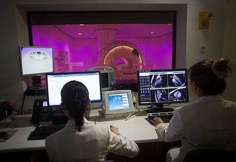
Magnetic Resonance Imaging, or MRI, is another diagnostic tool. An MRI can be taken of the brain – to investigate for tumours and in the event of a stroke – and of other parts of the body. An MRI scan uses magnetism to build up a picture of the inside of the body while CT relies on X-rays. Both modalities have their uses and each has its relevance depending on what is being investigated.
Source: The Business Times © Singapore Press Holdings Limited. Permission required for reproduction.
Stay Healthy the Easy Way
Get trusted health advice, offers and more.

Get the Health Buddy App
 Get it on Google Play