


GPs are often the first line of diagnosis for thyroid nodules. While most nodules are benign, when is further investigation needed? Through an in-depth case vignette, we share insights on what to look out for, how to investigate and manage, and when to refer.
INITIAL PRESENTATION AND EXAMINATION
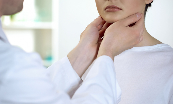
A 40-year-old lady with no past medical history presents with a two-week history of a right anterior neck lump. The mass has not increased in size and she has no other symptoms such as hoarseness or dysphagia.
What else would you do?
- History taking for risk factors for thyroid cancer
- Family history
- Exposure to radiation
- Check for symptoms of hyperthyroidism/hypothyroidism
- Examination
- Thyroid masses
- Cervical lymphadenopathy
- Check for red flags for thyroid cancer
- Rapid growth
- Persistent hoarseness suggesting recurrent laryngeal nerve involvement
- Dysphagia
- Hard/fixed mass
- Cervical lymphadenopathy
On further questioning, she notes that her mother had a history of thyroid cancer with surgery performed many years ago. She is clinically euthyroid. On examination, a 2 cm right thyroid nodule is palpated. This appears well circumscribed, firm and moves with swallowing. There is no cervical lymphadenopathy. She is very anxious about this nodule and asks if she has cancer.
What can you tell her?
- Thyroid nodules are very common and can occur in up to 60% of the population. This condition is more common in women.
- Majority (> 95%) of thyroid nodules are benign, while the remaining 5% are malignant1
- She does have a family history of thyroid cancer and would benefit from further workup of this nodule (5-10% of differentiated thyroid cancers have a familial occurrence1)
- However, given that only one first-degree family member is affected, she would not have benefitted from thyroid cancer screening with ultrasounds1
INVESTIGATIONS
She agrees to further workup. What investigations should be performed?
You can perform:
- Thyroid panel (thyroxine [T4] / thyroid-stimulating hormone [TSH])
It is recommended by the American Thyroid Association (ATA) that serum TSH be measured in the initial evaluation of a patient with a thyroid nodule. This is also relevant if the patient is subsequently planned for surgery as general anaesthesia can precipitate a thyroid storm in patients with uncontrolled hyperthyroidism. - Thyroid/neck ultrasound with survey of the cervical lymph nodes
It is preferable to have an experienced and accredited provider perform a formal diagnostic ultrasound of the neck as the pattern of sonographic features confers a risk of malignancy, and combined with nodule sizes, guides decision for fine needle aspiration (FNA).
Numerous systems provide guidelines on risk levels and decision making for FNA, including the ATA guidelines (Figure 1) as well as the American College of Radiologists Thyroid Imaging Reporting and Data System (ACR TI-RADS).
In Singapore General Hospital (SGH) and throughout SingHealth Polyclinics, the ACR TI-RADS system2 is employed for all thyroid ultrasound reports. Each ultrasound characteristic is given a value and the total number is then tallied to achieve a final score (ranging from TR 1-5). Based on this score, recommendations are given for FNA or follow-up.

WHEN TO REFER FOR SPECIALIST CAREYou decide to perform the thyroid panel and neck ultrasound and the results return as:
Based on these suspicious results, you refer her to the SGH Otorhinolaryngology - Head & Neck Surgery specialist outpatient clinic. What are other possible reasons for referral?
|
What patients can you manage without referral at this point?
- Purely cystic nodules without symptoms: risk
of malignancy < 1%. These nodules can also
be aspirated if you are comfortable with such
procedures, although aspiration carries the risk of bleeding, infection, re-accumulation and rarely
airway compromise
- Very low suspicious nodules (e.g., spongiform
nodules), which can be followed with serial
ultrasound (e.g., yearly)
- Nodules that have previously been investigated and have either two benign FNAs before or stable nodules with a previous benign FNA
WHAT TREATMENTS ARE AVAILABLE
The patient is seen in the SGH Otorhinolaryngology - Head & Neck Surgery specialist outpatient clinic and ultrasound guided FNA is performed. The results return as suspicious for papillary thyroid cancer and she is offered a hemithyroidectomy versus a total thyroidectomy.
What are the pros and cons of hemithyroidectomy versus total thyroidectomy?
Hemithyroidectomy
In tumours less than 4 cm, hemithyroidectomy has been found to have excellent survival in properly selected low- to intermediate-risk (with no extrathyroidal extension or lymph node metastases) patients. While there is a slightly higher risk of recurrence with hemithyroidectomies, salvage therapy is highly effective.
Total thyroidectomy
Total thyroidectomy carries increased risk of recurrent laryngeal nerve palsy and bilateral recurrent laryngeal nerve palsy (which may require a tracheostomy). Also, there is a risk of hypocalcaemia and there is a need for lifelong thyroxine replacement (as opposed to no risk of hypocalcaemia and 25% risk of requiring thyroxine supplementation for hemithyroidectomies).
The patient decides on a total thyroidectomy and enquires about minimally invasive options that she has found while doing an online search.
What can you tell her about this?
- Thyroidectomies (hemi and total) are mostly performed via a neck or transcervical skin incision. This affords the best access and visualisation of the thyroid gland and its surrounding structures such as the recurrent laryngeal nerves and parathyroid glands, but leaves a visible neck scar.
- For small thyroid lesions, a shortened neck incision may be adopted to remove the lesion via a video-assisted technique known as MIVAT (minimally-invasive video-assisted thyroidectomy). This method is, however, not suitable for all patients.
- Remote access approaches have been developed to avoid a neck scar. It is important to understand that fundamentally, these options do not offer an equivalent ease of access and they require a larger extent of dissection and longer operative time. These approaches site the scar away from the neck, and common locations include: post-auricular, axillary, anterior chest-wall/breast-axillary, and transoral. A combination of endoscopic and/or robotic instruments are usually required to perform the surgery.
These approaches are available at SGH but meticulous patient selection and careful risk-benefit consideration must be undertaken before embarking on surgery3.
POSTOPERATIVE CARE AFTER DISCHARGE
The patient undergoes a total thyroidectomy uneventfully. Postoperatively her calcium levels are normal and parathyroid hormone level is at the lower range of normal. By postoperative day (POD) three, her calcium levels are noted to be stable and she is discharged home with a surgical drain, with advice on how to care for the drain.
Scenario 1:
She presents to your clinic the day after discharge and her drain bottle has lost suction. What can you do?
- Assess the drainage amount
Each patient has been given a chart to record the drain amounts per 24 hours. This value is usually less than 50 ml / 24 hours by POD 3 and there should be a downtrend. The drainage should be serous or mildly haemoserous. - Assess for neck swelling
If there is no neck swelling, the drain suction can be re-created or drain bottle changed (each patient is usually provided with a new drain bottle). The patient should be sent to Accident & Emergency (A&E) if there is significant neck swelling, neck erythema/tenderness, purulent drainage, or shortness of breath.
Scenario 2:
She presents to your clinic the day after discharge complaining of perioral numbness and tingling in her hands and feet. What should you do?
- These are symptoms of hypocalcaemia
- Chvostek's sign (tapping over the parotid elicits twitching in the facial nerve) can be elicited but a negative sign is not sensitive and does not rule out hypocalcaemia
- The patient can be given a stat dose of 2.5 g calcium carbonate and 0.5 mcg Rocaltrol and an electrocardiogram (ECG) can be performed
- Unless able to send and check for calcium levels stat, the patient should be referred back to A&E for calcium level check and possible intravenous (IV) calcium replacement
FOLLOW-UP AND MONITORING
Histology results return as a 1.5 cm papillary thyroid carcinoma with no lymph node involvement. She is thus staged as a T1N0 papillary thyroid carcinoma and she is maintained on 100 mcg thyroxine.
How do we follow her up?
- Clinical follow-up and neck examination every 6-12 monthly.
- Thyroxine supplementation is used to maintain the TSH at a suppressed level, to reduce the risk of recurrence. The exact TSH target is dependent on factors including:
- Risk of recurrence
- Symptoms of hyperthyroidism
- Age
- Risk factors for hyperthyroidism (e.g., atrial fibrillation [AF
This may range from < 0.1 in high-risk patients to 0.5-2.0 mU/L in low-risk patients.
- Thyroglobulin (Tg) and thyroglobulin antibody (TgAb) levels. These are markers of differentiated thyroid cancer recurrence that are generally more useful only if a total thyroidectomy has been performed. These have more value when trended serially (i.e., an uprising trend suggests recurrent disease).
- Neck ultrasound. A scan may be performed 6-12 months from surgery to assess for any structural disease. Scans may also be ordered if there is clinical suspicion of disease (e.g., new neck mass or if there is an increase in Tg levels).
She remains well for 5 years with persistently low Tg levels and no evidence of disease on neck ultrasound. She is keen for discharge from the specialist clinic to be followed up under your care.
How can you monitor her?
- In low-risk patients that have an excellent response to therapy, the utility of subsequent Tg testing is not established. The time interval can be lengthened to at least 12-24 months.1
- Serum TSH should be measured at least every 12 months in all patients on thyroid replacement therapy, with the TSH goal 0.5-2.0 mU/L.
- On follow-up, neck examination should be performed to detect any masses.
- Patients should be referred back to specialist care if new head and neck symptoms develop, a neck mass is detected, or there is a rise in Tg.
REFERENCES
- Haugen BR et al. 2015 American Thyroid Association Management Guidelines for Adult patients with Thyroid Nodules and Differentiated Thyroid Cancer. Thyroid 2016;26(10)
- Franklin TN et al. ACR Thyroid Imaging, Reporting and Data System (TI-RADS): White Paper of the ACR TI-RADS Committee. JACR 2017;14(5):587-595
- Kiong KL et al. Transaxillary thyroidectomies: a comparative learning experience of robotic vs endoscopic thyroidectomies. Otolaryngol Head Neck Surg 2015;152(5):820-826
Dr Kimberley Kiong is a Consultant at the Singapore General Hospital Department of Otorhinolaryngology - Head & Neck Surgery, and the Sengkang General Hospital Department of Otolaryngology (ENT). She is also Consultant with the SingHealth Duke-NUS Head & Neck Centre, seeing general ear, nose and throat (ENT) cases as well as thyroid and head and neck tumours.
Dr Kiong completed her medical degree at the National University of Singapore, Yong Loo Lin School of Medicine in 2011 and finished her ENT specialist training under the SingHealth Residency Program in 2017. During this time, she has published several papers and received the Outstanding Resident Award on multiple occasions.
Dr Kiong has completed a prestigious advanced fellowship training at the MD Anderson Cancer Center in Houston, USA, and is currently specialised in Head and Neck Cancer Surgery. Her specific interests include endoscopic skull base surgery for tumours and transoral robotic surgery.
Dr Anna See graduated from the National University of Singapore (NUS), Yong Loo Lin School of Medicine in 2012. She completed her membership examination of the Royal College of Surgeons of Edinburgh (Surgery) in 2014, and obtained her Master of Medicine (ENT) from NUS in 2015. She became a certified ENT specialist in 2018 upon completion of her ENT residency training in Singapore.
Her main interests are in the treatment of head and neck cancers and thyroid lesions. Her practice also covers general adult ENT conditions. Apart from clinical medicine, she has a keen interest in research of head and neck cancers and has presented in various regional and international conferences.
GP Appointment Hotline: 6326 6060
Stay Healthy the Easy Way
Get trusted health advice, offers and more.

Get the Health Buddy App


Follow HealthXchange

