HealthXchange will NEVER ask you to transfer money over a call. If in doubt, call the 24/7 ScamShield helpline at 1799, or visit the ScamShield website at www.scamshield.gov.sg.
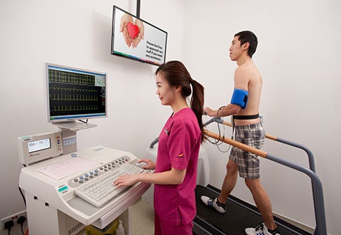
Patient using the exercise stress test (treadmill exercise).
Ever wonder how one goes through a diagnosis of a heart problem? A patient typically goes through one or more cardiac diagnostic tests to allow the doctor to provide an accurate prognosis of a heart disease and the appropriate treatment required.
A typical diagnosis starts from a physical examination and assessment of the patient and the patient’s family medical history. Based on the findings of the assessment, the doctor may request for selected cardiac diagnostic tests to be performed for the patient. Cardiac diagnostic tests are essential in the diagnosis of heart diseases such as coronary heart disease and arrhythmias (abnormal heart rhythms), and are usually carried out in a cardiac laboratory. Additional tests such as cardiac computed tomography (CT) scans may sometimes be carried out to detect the presence of narrowing in heart arteries in some patients.
The cardiac laboratory in NHCS provides a wide range of diagnostic tests and services that are designed to identify abnormalities in the function of one’s cardiac system. It is a full-fledged facility providing services to inpatients, outpatients as well as patients undergoing surgery in the operating theatre. The cardiac laboratory performs close to 45,000 echocardiograms (Echo) and 78,000 electrocardiograms (ECG) a year. The cardiac technologists are trained on-thejob at NHCS and accredited with allied health professional certification from American Registry of Diagnostic Medical Sonographers (Echo) and International Board of Heart Rhythm Examiners (Device Therapy/ Electrophysiology). They perform the non-invasive tests and generate preliminary reports for the cardiologists to decide on further invasive treatment.
Advancing Cardiac Diagnostic Tests’ Capability
A heart diagnostic device, the Spyder ECG, was recently mentioned by a minister on his Facebook, as he shared about how the Spyder ECG was used to monitor his heart rhythm.
The Spyder ECG is an innovative cardiac rhythm diagnostic solution developed by Assoc Prof Philip Wong, Senior Consultant of Department of Cardiology, NHCS and made in Singapore. Weighing only 49g, the Spyder ECG is a wireless wearable that can monitor one’s ECG rhythm continuously for up to 2 weeks. It is also used in patients with very short and infrequent symptoms such as palpitations, ‘missed beats’, fainting spells or irregular heartbeats, to increase the chance of capturing the abnormal rhythm.
The uniqueness of the Spyder ECG is that the ECG data is continuously transmitted and stored in a secured and centralised cloud database, thereby allowing medical technicians and doctors faster, real-time direct access to the ECG data. Unlike the traditional Holter Monitoring devices, where patients have to lug a recorder weighing 300g and put up with messy wires, the Spyder ECG is light-weight, inconspicuous, completely manageable by patients and even allows patients to view their own ECG on the paired smartphone.
Overall, the Spyder ECG system caters to an important disease area of cardiac arrhythmias; its compact size allows for increasing the duration of monitoring, and hence the likelihood of detecting abnormal rhythms such as Paroxysmal Atrial Fibrillation. Its fully digital and wireless data functionality allows quick turnaround time for reporting, shortening the diagnosis time needed for important treatments such anticoagulation for atrial fibrillation, pacemaker implants, or other medical therapy to be instituted efficiently.
Some Types of Cardiac Diagnostic Tests
Exercise Stress Test (Treadmill Exercise)
Exercise stress test allows the cardiologist to assess the response of the patient’s heart to the increased workload and demand for blood during exercise. It records the ECG of one’s heart while walking on a treadmill machine. This test is useful in diagnosing ischaemic heart disease (reduced blood supply to the heart muscles due to coronary artery disease).
Echocardiogram
Echocardiogram is a diagnostic procedure using high frequency sound waves (ultrasound) to take moving pictures of the heart and its related structures such as valves. The pictures or images appearing on the screen may be in black and white or colour.
From the pictures, the cardiologist can measure the size and function of heart chambers, study the motion of heart valves, and assess the efficacy of the contraction of heart muscles and the blood flow pattern across the valves and within the heart chambers. This allows the cardiologist to have better assessment on how well the heart is working and whether there are any abnormalities.
Upright Tilt Test
The upright tilt test is used to detect recurrent syncope (fainting) of unknown origin. A common form of syncope is called a vasovagal syncope, also refers to fainting due to abnormally sensitive reflexes in the cardiovascular system during prolonged standing or when subjected to unpleasant stimuli. Though this form of faints may appear alarming, they are rarely life-threatening. The test requires the patient to be tilted (70 degrees) in a standing position to simulate a situation of prolonged standing and he or she will be closely monitored for any symptoms of fainting.
Ambulatory ECG (Holter Monitoring)
The Ambulatory ECG - Holter Monitoring is a test where the ECG is continuously monitored for 24 or 48 hours and the signals are simultaneously recorded onto a special recorder worn by the patient, either at his or her own home or work environment. It allows any abnormal rhythms or ECG abnormalities to be captured during the monitoring period.
This test is useful for detecting transient rhythm disorders of the heart, which sometimes can be hard to detect. It is suitable for patients with palpitations, giddiness or fainting spells. By quantifying the amount and type of ECG abnormalities, it will be able to provide quantitative and qualitative information on the effect of the drug therapy.
The test can also help post-procedural patients, especially those who have undergone the electrophysiological studies, to determine if the procedure was successful.
Cardiac Computed Tomography (CT) Scan
The Cardiac CT scan is a non-invasive test which examines the coronary arteries and vessels that supply oxygenated blood to the heart wall. It is a relatively painless scan which allows doctors to obtain information about the location and the extent of narrowing and plaque in the coronary arteries with a high degree of accuracy.
Build-up of fats and other substances (known as plaque), can narrow the arteries or even close off blood flow to the heart over time. This may result in chest pain or a heart attack.
Two types of cardiac CT are available – CT coronary angiogram and coronary calcium scan. A CT coronary angiogram is primarily done to examine the coronary arteries by injecting a contrast dye into one of the arm veins. This allows our doctors to visualise the arteries and identify any narrowing in them.
A coronary calcium scan does not require contrast injection and provides information about the location and extent of calcium build-up in the coronary arteries, which is a useful indicator of the amount of cholesterol deposition (atherosclerosis).
This article is from Murmurs Issue 33 (Jan – Apr 2019). Click here to read the full issue.


 Get it on Google Play