


Major amputation of the lower limb is one of the most devastating complications of diabetes, and many cases begin as a foot ulcer. General practitioners play an important role through patient education, foot screening and the identification of ‘foot at risk’ patients for a timely referral to a tertiary centre, to reduce the risk of amputation.
INTRODUCTION
Diabetes mellitus is one of the most significant causes of ill health and disease in Singapore. The lifetime risk of developing diabetes is one in three among Singaporeans, and the number of those with diabetes is projected to surpass one million by 2050.1 This is associated with a projected increased expenditure in the care of these Singaporeans, which is estimated to rise to SGD 2.5 billion by 2050.2
DIABETES AND LOWER LIMB AMPUTATION
Major amputation of the lower limb is one of the most devastating complications of diabetes. Disability is common after amputation, and removing the individual from the workforce will cause further economic burden.
Foot ulceration due to neuropathy and repetitive trauma, complicated by infection and ischaemia, is the underlying pathway to amputation. The lifetime incidence of developing a foot ulcer may be as high as 25% in persons with diabetes3, and 84% of nontraumatic limb amputations are preceded by foot ulcers4.
Patients with diabetes have a tenfold higher risk of amputation compared to the general population.5 The prognosis after lower limb amputation is poor, with one-year mortality of up to 30% in Singapore.6
DIABETIC FOOT WOUND MANAGEMENT
The key to management of diabetic foot wounds is prevention, through education, foot screening and advice on appropriate foot care and footwear.
However, if a wound develops, it should be promptly referred to a tertiary centre for management by a multidisciplinary limb salvage team.
In 2009, Fitzgerald introduced the ‘toe and flow’ concept of the diabetic foot team.7 The ‘irreducible minimum’ of the team consists of a vascular surgeon and podiatrist, with frequent inclusion of additional specialists as available and necessary.
A systematic review by Musuuza et al. in 2020 reported a reduction in major amputation for diabetic foot wounds in 94% of the studies, and up to 51% absolute or 89% relative reduction in major amputation associated with management by multidisciplinary teams.8
The team compositions varied between studies, and included surgeons, physicians and allied health professionals. Four key clinical tasks commonly addressed were glycaemic control, local wound management, vascular disease and infection. The teams functioned in both inpatient and outpatient settings.
Multidisciplinary Care for Diabetic Foot Wounds at Changi General Hospital
The Changi General Hospital (CGH) Vascular and Endovascular Surgery Service has been involved in the care of diabetic foot wounds for many years. We have a specialised multidisciplinary team caring for these patients.
In the inpatient setting, the multidisciplinary team conducts ward rounds together every day to ensure optimal medical and wound care. For the past nine years, our service has had a consistent limb salvage rate of more than 90% at six months post-intervention.
THE CGH WOUND HEALING CENTRE
The CGH Wound Healing Centre provides a one-stop outpatient multidisciplinary service for the evaluation and management of chronic wounds, including diabetic foot wounds. The centre is helmed by board-accredited specialists from Vascular Surgery, Orthopaedic Surgery and Plastic Surgery.
Specialised wound care nurses conduct consultations, perform therapy for complex wounds and leverage a variety of technologies for wound management. Podiatrists perform gait analysis, biomechanical intervention and pressure offloading, and advise on proper foot care and footwear.
OUR FACILITIES AND CARE TEAM
There are facilities for bedside wound debridement (Figure 1) and advanced wound care such as the application of negative pressure wound therapy, ultrasound and electrical stimulation, and ultrasonic wound debridement (Figure 2). Diagnostic facilities such as ultrasonography for arterial disease and transcutaneous oxygen measurements are also available at our centre.
Our care team is certified by the American Board of Wound Management (ABWM) as Certified Wound Specialist Physicians (doctors) and Certified Wound Specialists (nurses and podiatrists).

Figure 1 The wound examination and dressing room has seven beds and ample space to allow for wheelchair access
OUR SERVICES
- Wound assessment
- Ultrasonography for arterial/venous pathology
- Transcutaneous oxygen measurement
- Conservative sharp wound debridement
- Simple/complex wound dressing
- Compression therapy for venous ulcers
- Negative pressure wound therapy
- Ultrasound and electrical stimulation for wound healing
- Ultrasonic-assisted wound debridement
- Podiatry for diabetic foot care

Figure 2 Advanced Practice Nurse Cheng Shuhua performing bedside ultrasonic wound debridement on a patient
CASE STUDY |
|---|
Background and presentation Mr A is a 63-year-old Indian male with a history of diabetes mellitus, hypertension and ischaemic heart disease. He is independent in his activities of daily living and is community ambulant with no aids. He presented to us with a poor-healing wound on his right heel associated with increasing swelling and pain (Figure 3). Figure 3 Infected right heel wound before and after initial debridement He was previously seen by a general practitioner (GP) and private vascular surgeon who advised him to go to a public institution for further management, as primary management with antibiotics and wound dressing did not improve the situation. He was not compliant with his diabetes medications. Evaluation On evaluation, Mr A was found to have right lower limb chronic limb-threatening ischaemia with an infected right heel wound and underlying calcaneal osteomyelitis. He also had poorly-controlled diabetes with a random fasting glucose of 17.2 mmol/L and HBA1c of 10.3%. Multidisciplinary management Mr A was reviewed by the endocrinologist for control of his diabetes and titration of his diabetes medications. He was also seen by the dietitian and diabetes nurse educator to reinforce his knowledge on diabetes control. Figure 4 A clean wound was achieved after another two surgical debridements with partial calcanectomy performed He underwent right lower limb angioplasty and debridement of the right heel wound. The infection was very extensive, involving the plantar fat pad and plantar fascia. He required another two surgical debridements and partial calcanectomy to achieve a clean wound, but the resultant wound was a large defect in the right heel (Figure 4). He was reviewed by the Plastic and Reconstructive Surgery team, and was offered the options of free flap reconstruction versus fish skin dermal substitute and negative pressure wound therapy. Mr A opted for the latter (Figure 5). Figure 5 Application of fish skin dermal substitute to right heel wound Mr A’s care was mainly led by the vascular surgeon, with contributions from the plastic surgeon, endocrinologist, infectious disease physician and allied health professionals including wound care nurses, podiatrists, physiotherapists and a diabetes nurse educator. Patient outcomes After several surgical procedures for wound debridement and application of fish skin dermal substitute, Mr A’s wound healed at seven months after his first operation (Figure 6). He maintains functional status and the ability to walk. Figure 6 From left to right: three, six and seven months after first surgery |


THE ROLE OF GPs IN DIABETIC FOOT CARE
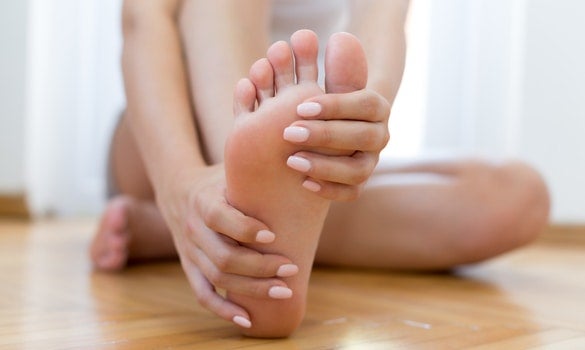
GPs play an important role in the prevention of diabetic foot ulcers through patient and caregiver education, foot screening, advice on foot care and footwear, and the identification of ‘foot at risk’ patients for referral to a tertiary centre for further evaluation.
Determining the risk of foot ulcers
The key risk factors for development of foot ulcerations are:
Loss of protective sensation (LOPS) / peripheral neuropathy
One or both distal pulses not being palpable / peripheral arterial disease (PAD)
Presence of foot deformity or callosity
The International Working Group of the Diabetic Foot (IWGDF) risk stratification system uses these three factors to advise on foot screening and examination frequency (Table 1).
Patient and caregiver education
Patient and caregiver education includes the following advice:9
Do not walk barefoot, in socks without shoes, or in thin-soled slippers, whether indoors or outdoors
Inspect daily the entire surface of both feet, and the insides of the shoes that will be worn
Wash feet daily with careful drying, especially in between toes
Use emollients to lubricate dry skin
Cut toenails straight across
Avoid using chemical agents, plasters or any other technique to remove calluses or corns
Self-monitor foot skin temperature to identify early signs of inflammation
Wear appropriate footwear that accommodates the shape of the feet and fits properly, to reduce plantar pressure and help prevent a foot ulcer
Category | Ulcer risk | Characteristics | Frequency |
|---|---|---|---|
0 | Very low | No LOPS and no PAD | Once a year |
1 | Low | LOPS or PAD | Once every 6-12 months |
2 | Moderate | LOPS + PAD, or OPS + foot deformity, or PAD + foot deformity | Once every 3-6 months |
3 | High | LOPS or PAD, and one or more of the following: • History of a foot ulcer • A lower-extremity amputation (minor or major) • End-stage renal disease | Once every 1-3 months |
Table 1 The IWGDF risk stratification system9
CONCLUSION
The key to decreasing the incidence of diabetic foot ulceration is prevention. If a foot ulcer develops, the patient should be referred to a tertiary centre for multidisciplinary management to reduce the risk of major amputation.
The CGH Wound Healing Centre is a multidisciplinary outpatient facility focusing on early intervention and the fast-track treatment of such patients. Together, we work towards one goal to improve limb salvage rates in diabetic patients, and to maintain their functional status and integrity.
REFERENCES
MOH. Diabetes: The war continues. 2017. https://www.moh.gov.sg/docs/librariesprovider5/war-on-diabetes/wod_public_report.pdf Accessed 6 December 2021
Png ME, Yoong J, Pham TP, Wee HL. Current and future economic burden of diabetes among working-age adults in Asia: conservative estimates for Singapore from 2010-2050. BMC Public Health. 2016;16:153.
Singh N, Armstrong DG, Lipsky BA.Preventing foot ulcers in patients with diabetes. JAMA. 2005;293:217-28.
Brem H, Sheehan P, Rosenberg HJ, Schneider JS, Boulton AJ. Evidence-based protocol for diabetic foot ulcers. Plat Reconstr Surg. 2006;117:7 Suppl193S-209S.
Siitonen OI, Niskanen LK, Laakso M, et al. Lower-extremity amputations in diabetic and nondiabetic patients. A population- based study in eastern Finland. Diabetes Care 1993; 16: 16–20.
Yee Ang, Chun Wei Yap, Nakul Saxena, Lee-Kai Lin, Bee Hoon Heng. Diabetes-related lower extremity amputations in Singapore. Proceedings of Singapore Healthcare 2017, Vol. 26(2) 76–80.
Fitzgerald RH, Mills JL, Joseph W, Armstrong DG. The diabetic rapid response acute foot team: 7 essential skills for targeted limb salvage. Eplasty. 2009; 9: e15
Jackson M, Bryn LS, Suleyman K, Prakash B, Christie MB, Meghan BB. A systematic review of multidisciplinary teams to reduce major amputations for patients with diabetic foot ulcers. J Vasc Surg. 2020;71(4):1433-1446.e3.
Bus SA, Lavery LA, Monteiro-
Soares M, et al. Guidelines on the prevention of foot ulcers in persons with diabetes (IWGDF 2019 update). Diabetes Metab Res Rev. 2020;36(S1):e3269. https://doi.org/10.1002/dmrr. 3269
Dr Lew Pei Shi is a Vascular and Endovascular Surgeon in the Department of General Surgery in Changi General Hospital (CGH). She has a special interest in the management of chronic and complex wounds and is an American Board of Wound Management Certified Wound Specialist Physician. She is part of the core group of surgeons heading the Wound Healing Centre in CGH.
GPs can call the SingHealth Duke-NUS Vascular Centre for appointments at the following hotlines, or scan the QR code for more information:
Singapore General Hospital: 6326 6060
Changi General Hospital: 6788 3003
Sengkang General Hospital: 6930 6000
KK Women’s and Children’s Hospital: 6692 2984
National Cancer Centre Singapore: 6436 8288
Stay Healthy the Easy Way
Get trusted health advice, offers and more.

Get the Health Buddy App


Follow HealthXchange

