HealthXchange will NEVER ask you to transfer money over a call. If in doubt, call the 24/7 ScamShield helpline at 1799, or visit the ScamShield website at www.scamshield.gov.sg.
Coming Soon: Precision Medicine for aggressive eye cancer
28 Jan 2020 | Inspire
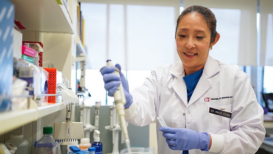
Frustrated by delays in diagnosing eye lymphoma, Dr Anita Chan decided to find a way to improve the process. The Clinician Scientist and Head, Ocular Inflammation & Immunology and Head, Translational Ophthalmic Pathology at the Singapore National Eye Centre, shares how her research has improved diagnosis of this disease and the role VisionSave has played in personalising Medicine in eye care.
What is eye lymphoma?
Eye lymphoma (vitreoretinal lymphoma) is an aggressive form of eye cancer. The symptoms include blurred vision and seeing multiple small dot-like shadows known as floaters. It is a subset of central nervous system lymphoma (CNS lymphoma), which is why up to 50-80 per cent of our patients go on to develop potentially fatal brain lesions. Early diagnosis and treatment of eye lymphoma not only saves the patient’s vision but also reduces the risk of lymphoma occurring in the brain.
Why was eye lymphoma difficult to diagnose and how has your research improved it?
To diagnose this disease, we need to confirm that cancerous cells are present in the eye. These cells float around in the vitreous, the jelly-like fluid inside the eyeball and need to be extracted by surgery or needle. It is difficult to diagnose because only a small volume of fluid can be removed from the eye and these cancer cells die very quickly once outside the eye. Previously, it would take many biopsies to confirm the diagnosis and by this time, the lymphoma would often have affected the brain. To address this, my team and I tested many methods to preserve the cancer cells for longer and found a specific “fixative” that would do so. This allows extra cancer cells to stay alive for us to now make a diagnosis with fewer biopsies and in a shorter time.
Once the disease is confirmed, patients are referred to the oncologists to check the brain and the rest of the body to find out the full extent of the lymphoma disease. Sometimes, the oncologist finds brain and other organs involved even before patients have symptoms, making the eye disease particularly important to diagnose.
Why is this research important for our local population?
Eye cancers are rare globally, which makes them difficult to study. The predominant form of eye cancer in Caucasians is uveal melanoma which affects the eye’s pigmented layer, the uvea. Therefore, in Europe and the United States, research has focused more on this disease than eye lymphoma. SNEC sees only about five to eight cases of eye lymphoma a year, but it is the most common form of cancer within the eye among Asians. Moreover, it’s associated with old age, so we can expect the number to rise with the ageing population.
Although the disease is rare, the impact is often devastating for patients and their loved ones because it can cause blindness, loss of body function and a change in mental state from the brain involvement. Keeping eye lymphoma cells alive for longer allows us to study the disease at the cellular and genomic level. This gives us a better understanding of the causes and disease pathways, and opens up the possibility of targeted personalised treatments to maximise outcomes.
How does a genomic understanding of eye lymphoma improve treatment?
The mutations that cause vitreoretinal lymphoma can vary and are not uniform across all of the lymphoma cells, even in the same patient. To get a true picture of a patient’s disease, we need to study each cell to identify the mutation present and proportion of these mutations such as homozygous (2 copies of mutation), heterozygous (one copy of mutation) or wild type (no mutations). This will allow us to separate individuals who clinically appear to have the same number of cancer cells in their eye lymphoma but actually may have more copies of the mutation (homozygous mutation) making the cancer more aggressive then someone who has only 1 copy of the mutation (heterozygous) or even no mutations. This is why two people diagnosed with similar eye lymphoma may still require different chemotherapy regimes. By paying attention to every single lymphoma cell, we can differentiate each person’s cancer and identify ways to target every single cancer cells in that person. This is what we mean by personalised therapy. In order for this research to succeed, we needed the support of SNEC and the Singapore Eye Research Institute’s (SERI) VisionSave initiative.
How did VisionSave help?
I needed a machine that selects out specific single cells and keeps them alive so they could be examined individually, but equipment funding can be difficult to secure for research. I discussed my proposed research project with the Executive director of SERI, Prof Aung Tin and he suggested that I apply for a VisionSave grant. I was fortunate to receive the funding in 2016 and established my laboratory within that year.
This research project also received the collaborative support of a biomedical company, Menarini Biomarkers Pte Ltd, that has helped to further fund the project. Together, we have filed a patent on this single cell approach as a diagnostic and prognostic test that identifies specific genetic mutations in eye lymphoma cells. Our collaborator aims to have the test licensed within the next two years so that it can be rolled out to help more people with this disease. The test will identify specific eye lymphoma mutations in a patient’s sample to allow oncologists to use this information to tailor treatments for the best outcomes. As a researcher, it is deeply satisfying knowing that such personalised eye care is now within reach and will have a direct impact on the health and lives of our patients.


 Get it on Google Play