HealthXchange will NEVER ask you to transfer money over a call. If in doubt, call the 24/7 ScamShield helpline at 1799, or visit the ScamShield website at www.scamshield.gov.sg.
One frequently asked question that heart doctors face is “How does coronary artery disease happen?” The main process behind the development of this disease is atherosclerosis.
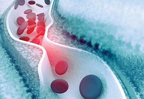
Atherosclerosis occurs over time with the deposition of plaque in the major blood vessels of the heart causing narrowing of the blood vessel and subsequently reduced blood flow to the heart muscles.
The plaque can consist of different types of materials including fats, fibrous tissue and calcium. The formation of plaque in the blood vessels is a result of a multitude of different factors and varies from person to person. These include age, gender, genetics, lifestyle, smoking and cardiac risk factors like diabetes, high blood pressure and high cholesterol. When the blockage is tight, common symptoms experienced include chest pain and shortness of breath, especially on exertion. This can also lead to heart attack.
Atherosclerosis - forming of plaque in the artery.
Treatment of coronary artery disease is multi-fold. Prevention, lifestyle modifications, control of risk factors and medications form the firm foundation of the treatment of this disease. With persistent symptoms despite adequate medical therapy, invasive treatment options can be considered. These include percutaneous coronary intervention for example, coronary stenting or coronary artery bypass (in the setting of multivessel disease).
Of the different types of blockages, calcified plaque poses a greater challenge for interventional cardiologists. Calcified blockages tend to be ‘rock-hard’ and as such, more difficult to ‘open up’ with the conventional equipment. Fortunately, the development and availability
of specialised technology, helps interventional cardiologists tackle these calcified lesions. The specialised devices used to treat these calcified blockages can be grouped largely into two groups – ‘balloons’ and ‘drills’.
Using Specialised ‘Balloon’ Technology
In routine cases of percutaneous coronary intervention, a wire is placed across the blocked artery and a small balloon is treaded over the wire to the area of blockage. The balloon is then dilated to ‘open up’ the blood vessel prior to placing a stent (metal scaffold to help keep the artery open). For calcified lesions that tend to be very hard, it may be difficult opening up the lesion using the normal balloon. The two types of specialised balloons available for calcified lesions are namely cutting balloon and intravascular lithotripsy (IVL) balloon. The cutting balloon is embedded with several tiny blades. When inflated, these tiny blades create microscopic cuts in the calcium allowing the calcium to break open and the blockages to be adequately dilated. The IVL balloon, developed in recent times, is based on the same lithotripsy concept of
using ultrasound waves to help breakdown kidney stones. The IVL balloon emits sound waves that will help modify and crack the calcium, allowing the calcified lesion to be more easily dilated thereafter.
The Use of ‘Drilling’ Devices
There are two main ‘drilling’ techniques that interventional cardiologists use to deal with complex lesions - rotablation and orbital atherectomy. While technical differences exist, in general these devices consist of a burr/crown that is coated with diamond. The diamond coating results in the device being ‘harder’ than calcium, allowing the calcium to be ‘shaved’ away when the device comes in contact with the calcium. After placing a specialised wire across the blockage, the device is activated over the wire to spin at high speeds (in the tens of thousands to hundreds of thousands revolutions per minute). This spinning action helps abrade away the inner areas of calcium.
Other techniques such as the use of laser, exist but these are currently not available in Singapore. The exact technique used to deal with calcified lesions will depend on the clinical scenario and may also involve a combination of various techniques. While calcified lesions used to pose much difficulties to the interventional cardiologists, the advent of these technologies have helped facilitate a smoother procedure with improved outcomes for the patients.
This article is from Murmurs Issue 37 (May – August 2020). Click here to read the full issue.


 Get it on Google Play