HealthXchange will NEVER ask you to transfer money over a call. If in doubt, call the 24/7 ScamShield helpline at 1799, or visit the ScamShield website at www.scamshield.gov.sg.
A Small Incision - Minimally Invasive Cardiac Surgery
31 Mar 2021 | Murmurs (NHCS)
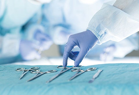
Traditionally, cardiac surgery is carried out through making a cut in the middle of the breastbone to gain access to the heart for maximal exposure. With technology advancement, minimally invasive cardiac surgery (MICS) can be carried out through a small incision (thoracotomy) on the side of the chest between ribs, without splitting the breastbone.
Peter Tan* is the sole breadwinner in his family. When he was first diagnosed with mitral valve disease, he was concerned that his condition would affect his work and was hesitant to undergo an open-heart surgery, as full recovery may take up to three months. Upon assessing Peter’s condition, Asst Prof Chua Kim Chai, Consultant from the Department of Cardiothoracic Surgery recommended minimally invasive mitral valve repair procedure where the recovery time is shorter. Peter underwent the procedure and was able to go back to work a few weeks after his surgery. He was happy that he had a short recovery time, and his symptoms have improved.
Wound healing is a much smoother and swift process with MICS.
“Compared to conventional cardiac surgery carried out through sternotomy, MICS has shown multiple benefits to patients such as minimising the need for blood transfusion, reducing the length of hospitalisation, hence saving medical costs. It also allows faster recovery and better cosmetic result due to less visible surgical scars,” shared Asst Prof Chua.
NHCS performed an average of 20 minimally invasive coronary bypass annually and had also just started performing minimally invasive mitral repair/replacement for suitable patients. Since October 2020, there had been 10 minimally invasive mitral repair/ replacement and the number is expected to increase with time. Each minimally invasive cardiac surgery depended on surgeon’s careful pre-operative planning and mastery of using long instruments, aided by using endoscope for visual improvement, to carry out the procedure. Prior to operation, patients also had to go through multiple preoperation investigations such as CT chest scan and lung function test.
Who are suitable for MICS?
MICS may not necessarily be an appropriate treatment option for every patient. Patients with serious medical condition such as lung disease or peripheral vascular disease, abnormal rib cage anatomy (severe kyphosis or pectus chest) and those require additional cardiac procedures, are not suitable to go for MICS. MICS is also generally not performed for complex cases that require multiple surgeries such as a combined valve and coronary artery bypass graft (CABG) surgery. MICS will bring greater benefit to patients who have an active lifestyle such as those who are into sporting activities, or those who are likely to have delayed chest bone healing if bone splitting was performed (osteoporosis or chronic steroid use).
It is important for patients to discuss with their doctors on the suitability for any treatment options, and undergo thorough assessment to determine the appropriate treatment option to achieve the best health outcome.
Selective Procedures Done Through MICS
Nowadays, selected coronary artery bypass graft (CABG) and valvular surgery can be done via minimally invasive approach. For CABG, a single graft bypass is most commonly performed on left anterior descending artery stenosis – minimally invasive direct coronary artery bypass, usually on a beating heart. For aortic valve replacement, either partial upper sternotomy, or right mini thoracotomy is the choice of exposure to the diseased valve. For mitral valve repair/replacement, sometimes with concomitant maze procedure (a surgical procedure used to treat atrial fibrillation) and/or tricuspid valve repair, the surgery is performed through right mini thoracotomy. Catheter-based procedures such as transcatheter aortic valve implant and mitral clip are part of MICS as well.
MICS is now a standard approach for straight forward cases in many parts of the world. In NHCS, with the well-equipped MICS equipment, trained anaesthetists, scrub nurses, perfusionists and experienced cardiologists, more of our patients could benefit from MICS in future.
* Patient’s name has been changed to protect his identity.
FOR PHYSICIANS: PROCEDURE IN-DEPTH
For minimally invasive direct coronary artery bypass, patient is positioned with slight right decubitus position and put on single right lung ventilation to allow better visualisation in left chest. A small incision of five to six centimetres is made on fourth intercostal space usually to gain exposure to target vessels. A specialised retractor is then applied through the incision to elevate left side of chest to allow harvesting of left internal mammary artery (LIMA). Then a suction device is used to stabilise left anterior descending (LAD) artery on a beating heart, and anastomosis of LIMA-LAD is performed thereafter.
For mitral valve surgery, the strategy of putting a patient on cardiopulmonary bypass is different from traditional way (centrally on ascending aorta and right atrium) – instead, it is established peripherally (femoral artery and vein). The access of heart is achieved through a four to five centimetres thoracotomy incision on right fourth intercostal space usually, aided with a thoracoscope for better visualisation of surgical field. Specialised long instruments are used for achieving diastolic arrest of heart, and subsequent repair or replacement of the diseased mitral valve.
This article is from Murmurs Issue 38 (September – December 2020). Click here to read the full issue.