


The Stress Echocardiogram captures images of the heart at its peak heart rate.
Dr Ewe See Hooi, Senior Consultant, Department of Cardiology, National Heart Centre Singapore (NHCS), a member of the SingHealth group shares two other common cardiac tests.
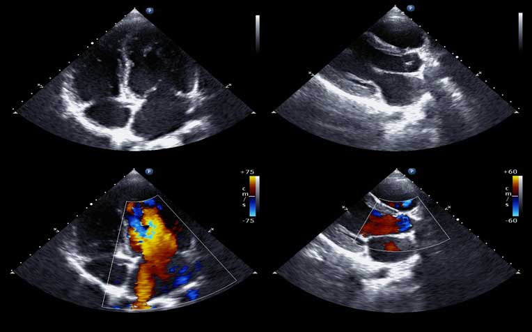
Stress echocardiogram
This test adds on to the stress test by including an imaging test precodure called echocardiography. Images of the heart are taken while the patient is performing the stress test or while lying down immediately after reaching peak exercise level. It captures images of the heart at its peak heart rate.
When is it done
- When the patient has chest pain.
- If the patient has had a previous heart attack, angioplasty or bypass surgery.
- If there is narrowing of the vessels supplying blood to the heart muscle.
- If there is an abnormal heart rhythm during exercise.
- As a screening test before starting an exercise programme or going for surgery.
- To assess symptoms and blood pressure response in patients with heart valve diseases.
Procedure
It uses ultrasound to study how the heart contracts and pumps blood while under stress. It also measures the size and function of the heart’s chambers, the motion of the heart valves, and the way blood is flowing. There are two kinds of stress tests. The pharmacological stress echocardiogram is for patients who are unable to exercise. The exercise stress echocardiogram is for those who can carry out treadmill exercises.
Pros and cons
A stress echocardiogram can identify the site of narrowed blood vessels, and the extent of reduced blood flow to the heart. There is a small possibility of the patient experiencing chest pain, irregular heartbeat, blood pressure changes, or a heart attack during the test. The doctor will take necessary precautions.
Nuclear cardiology test
In this test, images taken with a gamma camera can reveal which part of the heart is not getting supplied with enough blood and oxygen. It can also reveal the size of the affected area and the severity of the blockage.
When is it done
In more complex cases, such as when a patient has multiple blockages, or when a patient experiences chest pain after a previous surgery or angioplasty.
Procedure
It involves the injection of a radioactive tracer into the body, and use of a special gamma camera to track the tracer’s path. Radiographic images are then taken over a period of 15 to 20 minutes. The whole procedure takes two to three hours.
Pros and cons
It can reveal exactly where the heart is not getting enough blood and oxygen. But parts of the body near the heart, like the diaphragm or breast tissue, may produce some alterations that confuse the test results. The test does expose the patient to some radiation, but only within safe levels.
Ref: S13


Stay Healthy the Easy Way
Get trusted health advice, offers and more.

Get the Health Buddy App


Follow HealthXchange

