Bladder cancer may be divided into non-muscle invasive bladder cancer and muscle invasive bladder cancer. Doctors from the Urology Centre at Singapore General Hospital explains the diagnosis and treatments for bladder cancer.
Continued from previous page.
Diagnosis of bladder cancer
Bladder cancer can be diagnosed through the following clinical findings, laboratory and radiological tests, according to doctors from the
Urology Centre at
Singapore General Hospital, a member of the
SingHealth group.
-
Physical exam. The doctor feels the abdomen and pelvis for tumours and it may include a rectal or vaginal exam.
-
Urine tests. The laboratory checks the urine for blood, cancer cells and other signs of disease.
-
Ultrasound scan. This is a safe and painless test that uses sound waves to create images of organs and structures inside your body. It may be used to diagnose bladder cancer.
-
CT urogram or intravenous urogram (IVU). For patients with blood in the urine to visualise the whole urinary tract from the kidney to the bladder. A series of X-ray or CT scan images are taken after the dye is injected and secreted by the urinary tract.
-
Cystoscopy. A small tube, called a cystoscope, is passed through the opening of the urethra in the penis. It contains a lens and a light system that helps the doctor see the inside of the bladder lining to identify any tumours causing the bleeding. Tissue samples can be obtained from the bladder and tested for cancer cells.
Bladder cancer may be divided into:
- Non-muscle invasive bladder cancer (NMIBC), where the tumours are confined to the lining of the bladder.
- Muscle invasive bladder cancer, where the tumours have invaded into or beyond the muscular wall of the bladder.
The long-term outcome of bladder cancer depends on:
- Stage of the cancer, whether it is muscle-invasive or not
- Aggressiveness of the cancer under the microscope (grade)
- Bladder cancer cell type
- Patient’s age and general health
Treatment of bladder cancer
Non-muscle invasive bladder cancer
These cancers rarely spread and can usually be cured. Left untreated they may, in some cases, develop into muscle-invasive tumours.
These are usually treated in the following ways:
-
Transurethral resection of bladder tumour (TURBT). Under anaesthesia, an instrument called a resectoscope is inserted through the penis. The surgeon uses the resectoscope to remove the tumour tissue, one piece at a time, using a special wire loop. The pieces of tissue are flushed out at the end of the operation.
-
Intravesical therapy. After resection, chemotherapy agents such as mitomycin or immunotherapy such as BCG (Bacille Camete Guerin) therapy may be given through a catheter into the bladder to reduce the risk of recurrence and disease progression. Immunotherapy uses substances made by the body or in a laboratory to boost, direct, or restore the body's natural defenses against cancer.
Muscle-invasive bladder cancer
Muscle-invasive tumours have a high chance of spreading to other parts of the body and treatment is usually more aggressive.
Treatment options may include:
-
Surgery. Surgery involves removal of the entire bladder (radical cystectomy). Under general anaesthesia, the surgeon removes the entire urinary bladder and the surrounding lymph nodes in the pelvis. The prostate is removed in the male and the uterus, ovaries, fallopian tubes and part of the vagina are removed in the female.
-
Radiation therapy. Radiation therapy is a cancer treatment that uses high-energy X-rays to kill cancer cells or keep them from growing.
-
Chemotherapy. Chemotherapy uses anti-cancer drugs that kill cancer cells, or stop them from multiplying. It may be given before or after surgery. Patients may experience nausea, hair loss (alopecia), inflamed cheeks, gums, tongue, lips, and roof or floor of the mouth (stomatitis), and an abnormal blood profile that increases the risk of infection.
Sometimes a combination of treatment – chemotherapy with surgery or radiation – is needed to improve the chances of cure in selected patients.
Treatment under clinical trial
Photodynamic therapy (PDT) is a cancer treatment in which a light-sensitive drug is administered to the bladder and laser light is used to activate the drug to kill cancer cells.
What happens during surgery?
At the time of surgery, the entire bladder is removed. The ureters are disconnected from the bladder and joined to a loop of small intestines specially fashioned to contain urine. Depending on the pre-operative medical condition, stage of disease, and ability to perform clean intermittent self-catheterisation, the loop of small intestine may be:
- Connected directly to the abdominal wall and urine flows out through a urinary stoma (ileal conduit).
- Fashioned into a sphere (ileal neobladder) and reattached to the urethra. Urine passes out through the normal passage. Some patients may need to catheterise their urine passage regularly everyday to empty the bladder, as the neobladder does not have the sensory and contractile properties of the native bladder.
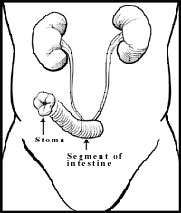
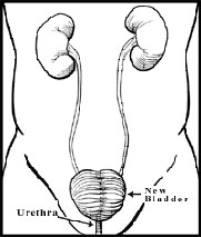
Figure 10
a. Ileal Conduit creation, where a segment of the intestine directs urine through a stoma into an external collecting bag,
b. ileal neobladder creation, where a loop of intestine is fashioned into a urine reservoir and connected to the urethra
See previous page for the types and
causes of bladder cancer.
Ref: N18
